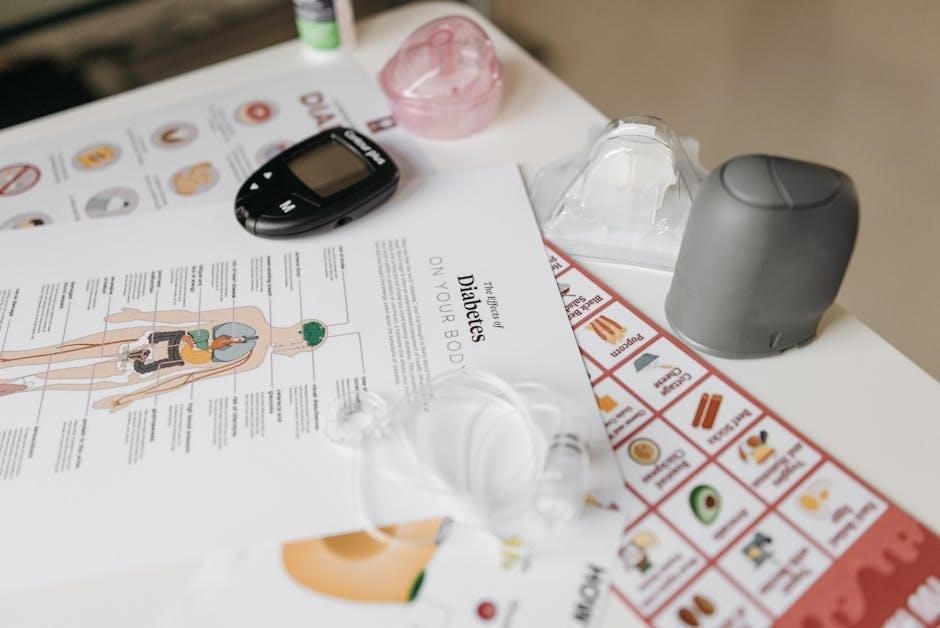
fluoroscopy guided injection
Fluoroscopy Guided Injection: A Comprehensive Overview (Updated 12/31/2025)
Today, December 31st, 2025, collaborative online tools like Microsoft Office enhance document creation and secure access to essential services, streamlining workflows effectively.
What is Fluoroscopy Guided Injection?
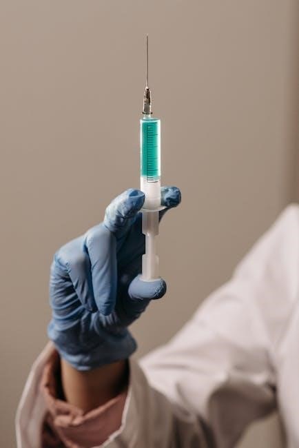
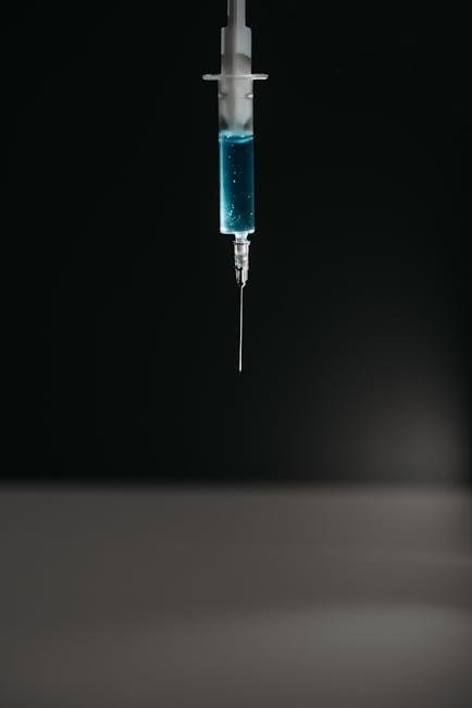
Fluoroscopy guided injection is a medical procedure utilizing real-time X-ray imaging – fluoroscopy – to precisely guide a needle to a specific location within the body. This technique allows physicians to visualize the needle’s path and ensure accurate medication delivery.
Unlike traditional “blind” injections, fluoroscopy offers a dynamic view, confirming the needle reaches the intended target – be it a joint, nerve, or soft tissue. This is particularly crucial for deep-seated structures or when anatomical landmarks are difficult to palpate.
The process involves injecting a contrast dye, visible under X-ray, to further confirm proper placement before administering the therapeutic medication. This method significantly improves the effectiveness and safety of various injection-based treatments.
The Role of Fluoroscopy in Injection Procedures
Fluoroscopy’s primary role in injection procedures is to provide real-time visualization, enhancing accuracy and minimizing risks. It transforms a static anatomical understanding into a dynamic, observable process. Physicians can directly witness the needle’s trajectory, avoiding critical structures like nerves and blood vessels.
The X-ray guidance allows for precise targeting, especially vital in complex areas like the spine or sacroiliac joints. Contrast dye injection, visualized under fluoroscopy, confirms correct placement before medication delivery. This ensures the therapeutic agent reaches the intended site, maximizing its effect.
Furthermore, fluoroscopy aids in verifying appropriate dispersion of the medication, optimizing treatment outcomes and reducing the need for repeat injections.
Why Use Fluoroscopic Guidance? — Accuracy & Safety
Fluoroscopic guidance dramatically improves injection accuracy, ensuring medication delivery to the precise anatomical location. This precision is paramount for maximizing therapeutic benefits and minimizing unwanted side effects. Safety is significantly enhanced by real-time visualization, allowing physicians to avoid critical structures.
Without fluoroscopy, injections rely heavily on anatomical landmarks, which can be imprecise. Fluoroscopy reduces the risk of nerve damage, vascular puncture, or unintended medication spread. The use of contrast dye further confirms correct needle placement before injecting medication.
Ultimately, fluoroscopic guidance translates to more effective pain relief and a reduced risk of complications, offering patients a safer and more reliable treatment experience.
Common Applications of Fluoroscopy Guided Injections
Fluoroscopy’s versatility enables precise injections targeting spinal structures, major joints, and soft tissues, offering diagnostic and therapeutic benefits across diverse musculoskeletal conditions.
Spinal Injections
Fluoroscopic guidance is paramount in spinal injection procedures, ensuring accurate needle placement within complex anatomical structures. This precision is crucial for delivering medication directly to the source of pain, maximizing therapeutic effects while minimizing risks.
Specifically, fluoroscopy allows visualization of vertebral levels, intervertebral foramina, and the epidural space. This real-time imaging capability is invaluable for procedures like Epidural Steroid Injections (ESIs), Selective Nerve Root Blocks, and Facet Joint Injections.
The use of contrast dye further enhances visualization, confirming appropriate medication distribution and identifying potential complications. Ultimately, fluoroscopy elevates the safety and efficacy of spinal injections, leading to improved patient outcomes and pain relief.
Epidural Steroid Injections (ESIs)
Fluoroscopically guided Epidural Steroid Injections (ESIs) are a cornerstone in managing spinal pain. Real-time X-ray imaging allows precise needle placement into the epidural space, the area surrounding the spinal cord. This accuracy is vital for delivering corticosteroids directly to inflamed nerve roots.
Contrast dye injection confirms proper placement and spread, ensuring the medication reaches the targeted area. Fluoroscopy minimizes the risk of dural puncture or vascular injury, enhancing patient safety. ESIs aim to reduce inflammation and alleviate pain associated with conditions like spinal stenosis, herniated discs, and sciatica.
The procedure’s success relies heavily on fluoroscopic visualization, optimizing medication delivery and maximizing pain relief potential for improved patient functionality.
Selective Nerve Root Blocks
Fluoroscopic guidance is paramount in performing Selective Nerve Root Blocks (SNRBs). These injections target specific nerve roots exiting the spinal column, pinpointing the source of radicular pain – pain radiating down an arm or leg. The real-time imaging allows physicians to meticulously navigate the needle alongside bony structures.
Contrast dye is crucial, confirming the medication’s precise location before injection. This minimizes the chance of non-target nerve blocks or unintended spread. SNRBs utilize local anesthetics, sometimes combined with corticosteroids, to temporarily interrupt pain signals.
Diagnostic and therapeutic, SNRBs help identify pain generators and provide temporary relief, aiding in treatment planning and potentially avoiding surgery.
Facet Joint Injections
Fluoroscopic guidance is essential for accurate placement of needles into the facet joints – small joints along the spine. These injections aim to alleviate pain stemming from facet joint arthritis or dysfunction, a common cause of chronic back pain. The live X-ray imaging ensures the needle reaches the targeted joint space.
Contrast dye injection confirms correct needle positioning, preventing unintended spread to surrounding nerves or tissues. Typically, a mixture of local anesthetic and a corticosteroid is delivered to reduce inflammation and numb the pain.
Facet joint injections offer both diagnostic and therapeutic benefits, helping pinpoint the source of pain and providing temporary relief, potentially delaying or avoiding surgical intervention.
Joint Injections
Fluoroscopy significantly enhances the precision of joint injections, ensuring medication delivery directly into the affected area. This technique is widely utilized for various joints, including the knee, hip, and shoulder, offering targeted pain relief and improved function. Real-time imaging allows physicians to visualize needle placement, minimizing the risk of complications.
The procedure typically involves injecting a corticosteroid and local anesthetic mixture to reduce inflammation and provide immediate pain relief. Fluoroscopic guidance confirms accurate placement within the joint capsule, maximizing therapeutic effect.
Joint injections are valuable for managing osteoarthritis, bursitis, and other joint-related conditions, improving patient quality of life.
Knee Injections
Fluoroscopically guided knee injections are a cornerstone in managing osteoarthritis and other knee joint pathologies. The real-time X-ray guidance allows precise needle placement within the joint space, ensuring the medication reaches the targeted area for optimal therapeutic benefit. This minimizes the risk of injecting into surrounding tissues.
Typically, a corticosteroid combined with a local anesthetic is injected, providing both immediate pain relief and longer-term anti-inflammatory effects. Fluoroscopy confirms accurate dispersion of the medication throughout the knee joint.
Patients often experience reduced pain and improved mobility following the procedure, enhancing their ability to participate in daily activities.
Hip Injections
Fluoroscopic guidance is invaluable for hip injections, due to the joint’s deep location and complex anatomy. Precise needle placement is crucial to deliver medication directly into the hip joint capsule, maximizing efficacy and minimizing complications. Without guidance, achieving accurate placement can be challenging.
These injections commonly utilize corticosteroids and local anesthetics to alleviate pain associated with osteoarthritis, bursitis, or labral tears. Fluoroscopy allows the physician to visualize the needle’s trajectory in real-time, confirming correct positioning before injecting.
Patients may experience significant pain reduction and improved range of motion, facilitating a return to functional activities.
Shoulder Injections
Fluoroscopically guided shoulder injections enhance precision when targeting the glenohumeral joint, a complex structure prone to various pain conditions. Accurate needle placement is vital for delivering therapeutic agents directly to the source of discomfort, improving treatment outcomes.
These injections often involve corticosteroids combined with local anesthetics, addressing pain stemming from rotator cuff tendinitis, bursitis, or osteoarthritis. Real-time visualization via fluoroscopy confirms the needle’s position within the joint space, avoiding unintended tissue contact.
Successful injections can significantly reduce pain, restore shoulder function, and improve the patient’s quality of life, enabling greater participation in daily activities.
Soft Tissue Injections
Fluoroscopic guidance extends beyond bony structures, proving invaluable for precise soft tissue injections. Targeting trigger points or inflamed bursae requires accurate needle placement to maximize therapeutic effect and minimize complications.
For trigger point injections, fluoroscopy confirms the needle tip resides within the taut band of muscle, delivering local anesthetic and potentially corticosteroids to alleviate myofascial pain. Similarly, in bursitis, visualization ensures medication reaches the inflamed bursa.
This guidance is particularly helpful in areas with complex anatomy or when palpation is unreliable. Utilizing real-time imaging enhances safety and efficacy, leading to improved pain relief and functional restoration.
Trigger Point Injections
Fluoroscopically guided trigger point injections offer a precise method for addressing myofascial pain syndromes. Real-time visualization allows physicians to accurately target the hyperirritable spot within the muscle’s taut band, ensuring the medication—typically a local anesthetic, sometimes combined with a corticosteroid—is delivered directly to the source of pain.
This accuracy is crucial as it minimizes the risk of injecting into surrounding structures and maximizes the therapeutic effect. The contrast dye can confirm proper needle placement, verifying the spread of the injectate within the targeted muscle tissue.
Improved outcomes and reduced discomfort are often reported with fluoroscopic guidance, especially in cases of deep or difficult-to-palpate trigger points.
Bursitis Injections
Fluoroscopic guidance significantly enhances the precision of bursitis injections, particularly around complex joints like the shoulder, hip, or elbow. Identifying the inflamed bursa can be challenging through palpation alone; fluoroscopy provides real-time visualization, confirming accurate needle placement within the bursal sac.
Contrast dye injection allows physicians to observe the spread of the medication, ensuring it reaches the intended target and avoids unintended injection into adjacent tissues or vessels. This is especially important in areas with numerous anatomical structures.
The use of fluoroscopy minimizes the risk of complications and maximizes the therapeutic benefit of corticosteroids or local anesthetics, leading to improved pain relief and functional outcomes.
The Fluoroscopy Procedure: A Step-by-Step Guide
Efficiently utilizing online Microsoft tools, the procedure involves careful preparation, sterile technique, precise needle placement visualized by fluoroscopy, and controlled injection delivery.
Patient Preparation
Prior to the fluoroscopy guided injection, comprehensive patient preparation is crucial for a successful and safe procedure. This begins with a thorough review of the patient’s medical history, including allergies, current medications, and any pre-existing conditions. Patients are typically asked to refrain from eating or drinking for a specified period before the injection, often several hours, to minimize the risk of complications.
It’s essential to inform the patient about the procedure itself, explaining the use of fluoroscopy, the sensations they might experience, and potential risks. Patients should also be encouraged to ask questions and voice any concerns they may have. Finally, ensuring the patient understands and signs a consent form is a vital step, confirming their agreement to proceed with the injection.
Sterile Technique & Skin Preparation
Maintaining strict sterile technique is paramount throughout the fluoroscopy guided injection procedure to prevent infection. The interventional radiologist and assisting staff meticulously scrub their hands and arms, donning sterile gowns, gloves, and masks. The patient’s skin at the injection site undergoes a rigorous preparation process, beginning with a cleansing solution to remove any surface bacteria.
Following cleansing, a broad-spectrum antiseptic, such as chlorhexidine or povidone-iodine, is applied to the skin and allowed to dry completely. Sterile drapes are then carefully applied to create a sterile field around the injection site, isolating it from potential contaminants. This meticulous approach minimizes the risk of introducing bacteria into the body during the procedure.
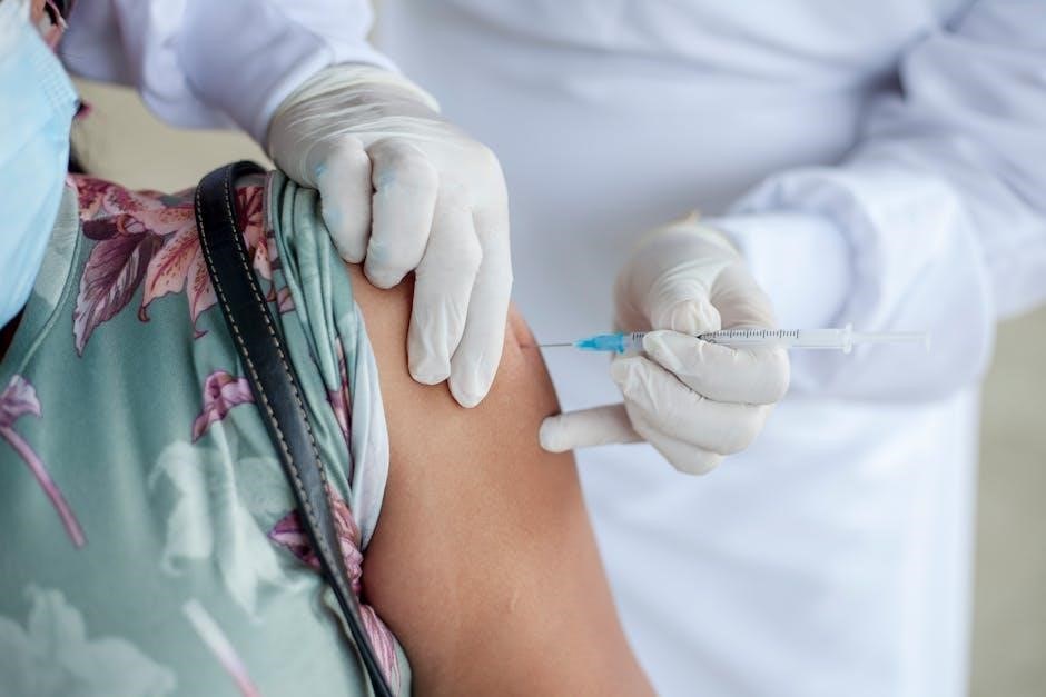
Needle Placement & Contrast Injection
With the patient positioned and the sterile field established, the radiologist carefully introduces the injection needle towards the target area, guided by real-time fluoroscopic imaging. Precise needle placement is crucial for accurate medication delivery and minimizing complications. Once the needle is positioned appropriately, a small amount of contrast dye – a radiopaque substance – is injected.
This contrast injection allows the radiologist to visualize the needle’s trajectory and confirm its location within the intended anatomical structure. The spread of the contrast dye helps assess the target area and ensure the medication will reach the desired tissues. This step is vital for verifying accuracy before administering the therapeutic agent.
Real-Time Imaging & Injection Delivery
Continuous fluoroscopic imaging is maintained throughout the injection process, providing a dynamic view of the needle’s position and the medication’s dispersion. The radiologist observes the contrast dye’s flow, confirming it reaches the intended target – be it a nerve root, joint space, or soft tissue. Once optimal placement is verified, the therapeutic agent, such as a steroid or local anesthetic, is slowly injected.
The injection rate is carefully controlled to minimize pressure and maximize drug efficacy. Real-time visualization ensures the medication is distributed effectively, avoiding unintended spread to surrounding structures. Throughout delivery, the patient is monitored for any discomfort or adverse reactions, ensuring a safe and precise procedure.
Risks and Complications of Fluoroscopy Guided Injections
Potential risks include radiation exposure, infection, bleeding, nerve damage, and allergic reactions to contrast dye; careful technique minimizes these possibilities effectively.
Radiation Exposure
Fluoroscopy utilizes ionizing radiation to create real-time X-ray images, guiding precise needle placement during injections. While essential for accuracy, radiation exposure is a valid concern. However, modern fluoroscopy units employ techniques to minimize dosage, such as pulsed fluoroscopy and collimation, focusing the beam only on the area of interest.
The amount of radiation received during a single fluoroscopy-guided injection is generally considered low, comparable to background radiation exposure over several months. Nevertheless, healthcare professionals adhere to the ALARA principle – “As Low As Reasonably Achievable” – to further reduce risk. Patient factors, like weight and the number of images required, influence the total dose.
Shielding, including lead aprons and thyroid shields, is routinely used to protect vulnerable body parts. Detailed records of radiation exposure are maintained for each patient, ensuring responsible and monitored use of this valuable diagnostic tool.
Infection
Although rare, infection is a potential complication of any injection procedure, including those guided by fluoroscopy. The risk arises from introducing bacteria into the body during needle insertion. Strict adherence to sterile technique is paramount in minimizing this possibility.
Healthcare professionals meticulously prepare the skin with antiseptic solutions, like chlorhexidine or iodine, before the injection. Sterile gloves, gowns, and drapes are consistently used throughout the procedure. The needle itself is single-use and disposed of properly after each injection.
Post-injection, patients are advised to monitor for signs of infection, such as increasing pain, redness, swelling, warmth, or fever; Prompt medical attention is crucial if any of these symptoms develop, allowing for timely diagnosis and treatment with antibiotics.
Bleeding & Hematoma Formation
Bleeding and hematoma formation are relatively common, yet usually minor, complications following fluoroscopy-guided injections. These occur when blood vessels are punctured during needle insertion. Individuals on blood thinners, or with bleeding disorders, are at a higher risk.
Typically, the bleeding is minimal and self-limiting, resolving within a few days. However, a hematoma – a collection of blood under the skin – can develop, causing localized pain, swelling, and bruising. Applying ice to the injection site immediately after the procedure can help minimize bleeding.
Patients are instructed to avoid strenuous activity and blood-thinning medications (when medically safe) for a short period post-injection. Larger hematomas may require medical evaluation and, rarely, drainage;
Nerve Damage
Although rare, nerve damage is a potential, though serious, complication of fluoroscopy-guided injections. This risk arises from direct needle trauma to a nerve or from subsequent compression due to hematoma formation. Symptoms can range from a transient tingling or numbness to more persistent pain, weakness, or even paralysis.
Precise needle placement guided by fluoroscopy significantly minimizes this risk, as does careful technique by the physician. However, anatomical variations and unexpected patient movements can contribute.
If nerve damage is suspected, immediate medical evaluation is crucial. Treatment may involve medication to reduce inflammation, physical therapy, or, in severe cases, surgical intervention. Complete recovery isn’t always guaranteed.
Allergic Reaction to Contrast Dye
Contrast dye, used to visualize structures during fluoroscopy, can occasionally trigger allergic reactions. These reactions vary in severity, ranging from mild itching and hives to a life-threatening anaphylactic response. Patients with known allergies to iodine or shellfish are at a higher risk.
Prior to injection, physicians routinely inquire about allergy history. Pre-medication with antihistamines or corticosteroids may be administered to mitigate potential reactions. During the procedure, staff closely monitor for any signs of allergy.
Symptoms of a reaction include difficulty breathing, swelling of the face or throat, and dizziness. Immediate medical intervention is vital if a severe reaction occurs, including epinephrine administration.
Alternatives to Fluoroscopy Guidance
Modern imaging offers alternatives like ultrasound, CT, and MRI for injection guidance, providing visualization without ionizing radiation, enhancing precision and patient safety.
Ultrasound Guidance
Ultrasound guidance utilizes high-frequency sound waves to create real-time images of soft tissues and structures. This technique is particularly valuable for visualizing tendons, ligaments, muscles, and superficial joints, making it ideal for procedures like soft tissue injections and some joint aspirations.
Unlike fluoroscopy, ultrasound does not involve ionizing radiation, presenting a significant advantage for patients concerned about radiation exposure. It’s also relatively inexpensive and portable, allowing for bedside procedures. However, ultrasound’s penetration depth is limited, making it less effective for visualizing deep structures like the spine.
The skill of the operator is crucial for accurate needle placement with ultrasound guidance, requiring extensive training and experience in musculoskeletal imaging. It’s a dynamic technique, allowing for adjustments during the injection based on real-time visualization.
CT Guidance
Computed Tomography (CT) guidance employs X-rays to generate detailed cross-sectional images of the body, offering superior visualization of bony structures and deep tissues compared to fluoroscopy or ultrasound. This makes CT particularly useful for guiding injections into the spine, sacroiliac joints, and other areas where bone obscures the target.
While providing excellent anatomical detail, CT guidance involves a higher dose of ionizing radiation than fluoroscopy. Therefore, it’s generally reserved for cases where fluoroscopy is insufficient or contraindicated. The procedure typically requires the patient to lie still within the CT scanner during image acquisition and injection.

CT’s ability to visualize both soft and hard tissues allows for precise needle placement, minimizing the risk of misdirection and maximizing therapeutic efficacy.
MRI Guidance
Magnetic Resonance Imaging (MRI) guidance utilizes strong magnetic fields and radio waves to create highly detailed images of soft tissues, ligaments, tendons, and nerves. Unlike fluoroscopy or CT scans, MRI doesn’t employ ionizing radiation, making it a potentially safer option for certain patients, particularly those requiring repeated imaging.
However, MRI guidance is less commonly used for real-time injection procedures due to the time required for image acquisition and the limitations of maneuvering instruments within the MRI scanner. It’s often employed for pre-procedural planning to precisely identify anatomical targets.
MRI’s exceptional soft tissue contrast is invaluable for visualizing subtle anatomical variations and guiding injections into areas like the hip or shoulder joint.
Post-Injection Care & Recovery
Following injection, adhere to activity restrictions, manage pain with prescribed methods, and promptly report any unusual side effects to your healthcare provider.
Activity Restrictions
Post-fluoroscopy guided injection, adhering to specific activity restrictions is crucial for optimal healing and maximizing the procedure’s benefits. Generally, patients are advised to avoid strenuous activities, heavy lifting (typically over 10-15 pounds), and vigorous exercise for a period ranging from 24 hours to several weeks.
The duration of these restrictions depends on the injection site, the specific condition treated, and the individual patient’s response. Your physician will provide tailored instructions regarding resuming normal activities, including work, driving, and recreational pursuits.
It’s important to gradually increase activity levels, avoiding sudden movements or overexertion. Ignoring these guidelines could potentially lead to complications or diminish the effectiveness of the injection.
Pain Management
Following a fluoroscopy guided injection, experiencing some mild discomfort at the injection site is common. This typically resolves within a few days and can be managed with over-the-counter pain relievers like acetaminophen or ibuprofen, as directed by your physician.
Avoid aspirin or other blood-thinning medications unless specifically approved, as they may increase the risk of bleeding. Applying ice packs to the injection site for 15-20 minutes at a time, several times a day, can also help reduce pain and swelling.
If pain persists or worsens despite these measures, contact your healthcare provider. They may recommend additional pain management strategies or investigate potential complications.
Potential Side Effects & When to Seek Medical Attention
While generally safe, fluoroscopy guided injections can cause temporary side effects like soreness, bruising, or mild pain at the injection site. More serious, though rare, complications require immediate medical attention.

Seek immediate care if you experience: fever, chills, increasing redness or swelling, severe pain, signs of infection (pus or drainage), numbness or weakness in the affected limb, or any allergic reaction (hives, difficulty breathing).
Contact your doctor promptly for any concerning symptoms that develop after the procedure. Early intervention can prevent minor issues from escalating into more significant problems, ensuring optimal recovery.
Leave a Reply
You must be logged in to post a comment.